
LASIK and higher order aberrations
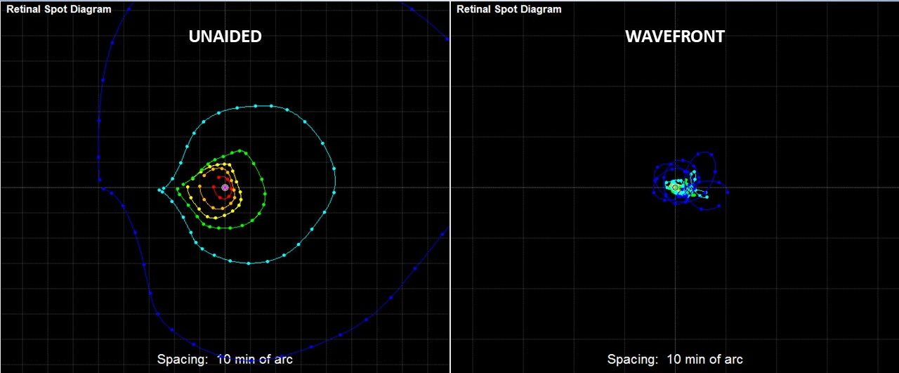
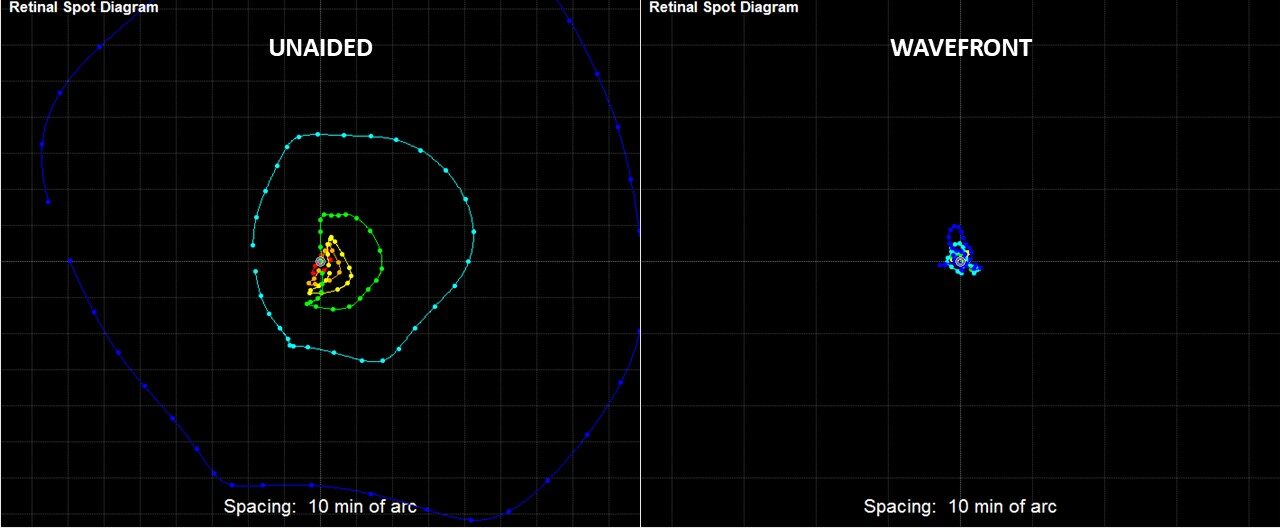
Most higher order aberrations in nature are what we would call PUPIL dependent. This means that they increase with increasing pupil diameter. When dealing with optical lenses, then we refer to it as APERTURE dependent. Below are scans from a ray-tracing aberrometer (iTrace, Tracey Technologies). The colored dots indicate individual rays of light projected by a laser through the pupil and traced to the point on the retina where each one lands. This is called a retinal spot diagram. The right eye is represented by the scans on the top and the left eye is below. All scans were obtained through dilated pupils. On the left side are scans of the unaided eye, meaning they were obtained without wearing contact lenses or any kind of correction. The unaided scans are very widely spaced, and the outermost rings are more widely spaced than the innermost. On the right side, these are the scans taken through LASERFIT wavefront correcting scleral lenses, and the differences are remarkable. Now the right eye did not correct as well as the left eye in Mathew’s case. But, it is nevertheless a very good scan, well-within the normal range. But Mathew had LASIK. Wouldn’t you expect those scans to be as tight as those through the special lenses? We’re going to dig a little deeper and find out why Mathew travels from the EU to LASERFIT each year. To be honest, we are not using his real name as we want to protect his identity.
What IS normal?
Normal is defined statistically. Before 1994, there was not even an instrument that could measure higher order aberrations. By the way, what do we mean by “higher order aberrations”? Simply stated, they are the optical imperfections in a system which are not correctable by standard lenses used in contact lenses and spectacles. What happened in 1994? It was the year when Junzhong Liang and colleagues invented the first instrument to measure human aberrations at the University of Heidelberg. Shortly after, Dr. Liang would publish the first articles on human higher order aberrations with David Williams at the University of Rochester. So, now that a way to measure these things exists, it required going into the field and measuring real eyes. We tend to use the study titled Normal-eye Zernike coefficients and root-mean-square wavefront errors by Thomas O. Salmon, OD, PhD, et. al., the largest multi-center study on thousands of normal eyes and normalized for pupil sizes at 3.0, 4.0, 5.0 and 6.0 mm. For larger pupils, then we have to use a different study titled High-order aberrations and preoperative associated factors by Luis E. Fernandez de Castro, et. al., which goes up to 7.0 mm. Both have similar mean values for each of their respective pupil diameters. So, when I tell a new patient that their uncorrected higher order aberrations with dilated pupils are multiple times higher than an average person who has not had surgery, this is the comparison I am making. That patient could very well have had much higher-than-average values prior to LASIK, or much lower, in which case that would be the baseline value for that person. Since we do not have access to that data, then I use the comparison with population norms.
Has anything changed?
Good question. Because Dr. Gemoules published a paper in 2007 on the higher order aberrations of a sample of patients who had refractive surgery (LASIK, PRK, or other) before and after fitting with special lenses, it is possible to go back to that time and make a rough comparison to today. The title of that paper is Rigid Gas-Permeable Contact Lenses and Severe Higher-Order Aberrations in Postsurgical corneas by Gemoules and Morris. The mean pupil diameter of the sample was 6.12 mm, and the mean higher-order root-mean-square TOTAL RMS – or HORMS – was 1.009 microns. The corresponding value for a 6.0 mm pupil size taken from de Castro is 0.389 microns, which means that for a pupil-equivalent basis, the unaided HORMS result was from 2 to 3 times greater than the pre-surgery group. We compiled another group in 2011 which had a mean pupil diameter of 5.9 mm and a mean HORMS of about 0.5 microns – or about half of what might be expected based on the earlier group from 2007. It appears to us as if maybe some changes were incorporated in the LASIK procedure itself which might have made a significant positive impact on the HORMS of the post-LASIK patients, and this change mostly applied to pupil sizes at or below 6.0 mm as we shall see in Mathew’s case.
But how does the patient see?
You may think that the aberrometer does a great job of quantifying the problem, but how does that relate to what the patient sees? What the aberrometer sees is the optical quality of the eye, which is the result of light entering the pupil and passing through the main refracting surfaces of the cornea and the crystalline lens (anterior aqueous and posterior vitreous to be precise). But first the light passes through the tear film, and that is often overlooked. What the patient sees is a result of the optics of the eye which then passes along the optic nerve and its various retinal layers and ganglia until it reaches the visual cortex in the posterior part of the brain. Does LASIK affect the neurological aspects of vision? Doubtful, but it certainly does alter the optics of the eye. Therefore, as vision specialists, we are actually mainly concerned about the quality of the optics. On our aberrometer, the iTrace, we can convert the retinal spot diagram into a Snellen letter (either a “C” or more commonly the “E”). This is a critical step in establishing a common objective reference by showing that image to the patient and asking them, “is that what you see”? Most of the time they say yes. So, then we can proceed to work on improving the sharpness of that image using a common reference.
Visual quality as a function of pupil diameter
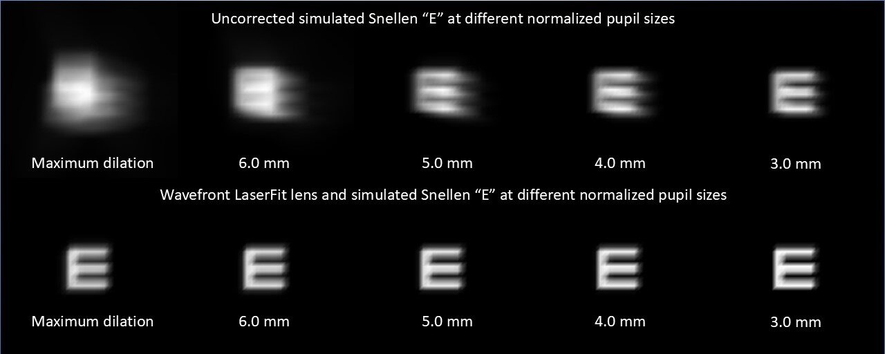
The above graphic compares the left eye without any lens (top) and with the final LASERFIT wavefront lens (bottom). These images were acquired by maximally dilating the pupil with 0.5% tropicamide and then normalizing the pupil at various standard diameters to show how the effects not only of the scleral lens, but also the effects of pupil diameter. In the case of the uncorrected post-LASIK eye, the image appears to be blurry at all pupil sizes, but certainly much clearer with the 3.0 mm pupil than the maximally dilated one. However, and this is a remarkable property of our LASERFIT process, almost all of the pupil diameters demonstrate good image quality and all seem clear.
Pupil size is a dynamic function depending on ambient lighting as well as the age and emotional state of the patient. Traditionally the unhappy LASIK patient complains about image quality at night or dim illumination. The age is typically from mid-20’s to mid-30’s, during that point in one’s life when the patient is active socially. Mathew was a university student when we first saw him. In other words, since pupil size is a continually changing variable, what should be our reference size for correcting the aberrations? Should it be like how some studies report their results at 5.0 mm, or should it be at the maximal dilation as might occur on a dark night driving on an interstate highway? We choose the latter and that is why this graphic shows sharp letters at all pupil sizes starting with the maximum diameter.
Putting it all together
The analysis
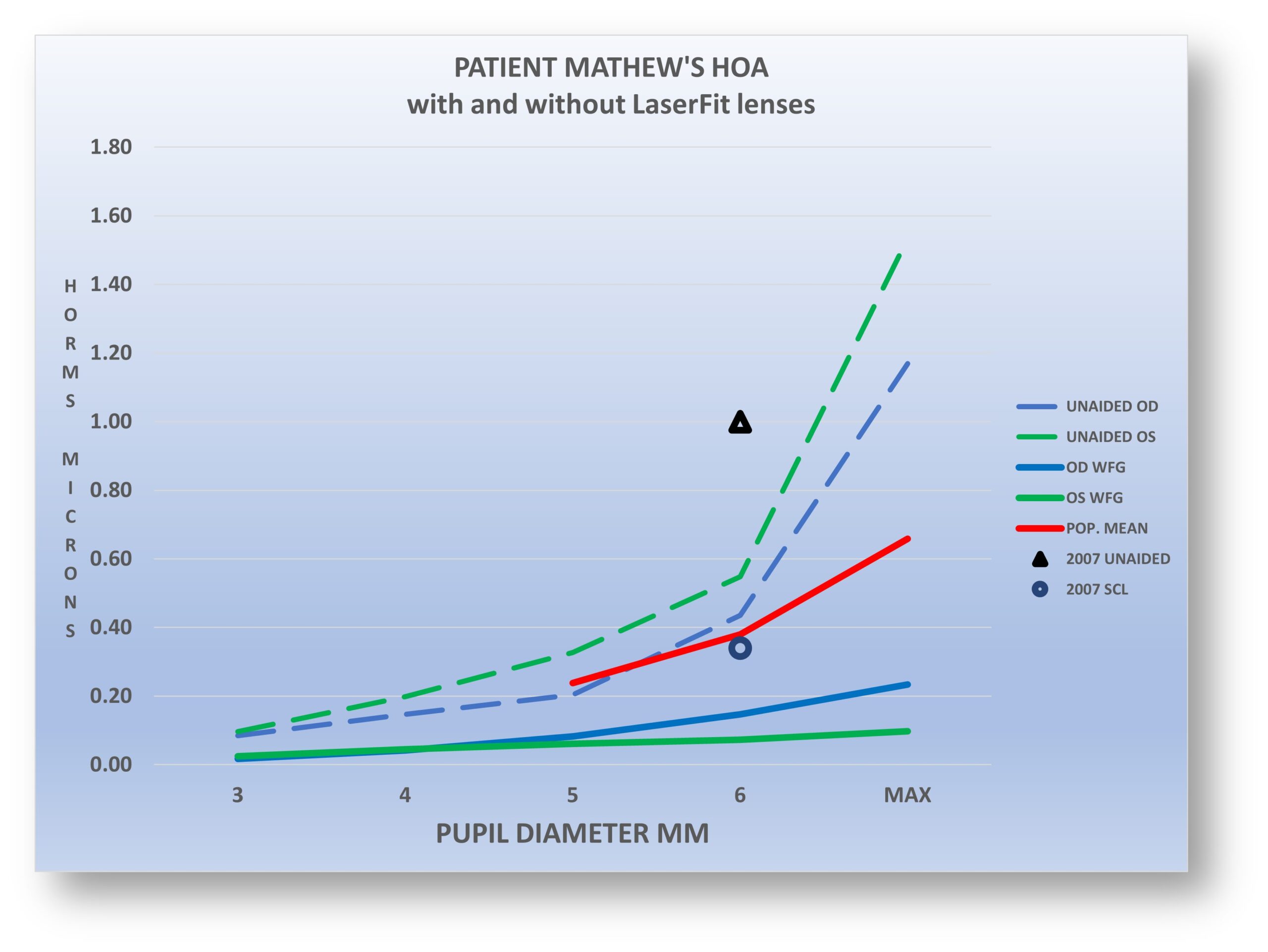
Here is the whole enchilada, showing the HORMS for both eyes with and without lenses and showing the population mean values in RED, and the 2007 reference point for a 6.0 mm pupil. Let’s examine the uncorrected HORMS for both eyes (dotted lines) and we can see that the values are fairly stable up until the 6.0 mm pupil diameter. This is now what we expect to see for many LASIK patients: a comparatively mild increase in HORMS up to about 6.5 mm, after which the values increase dramatically. Now look at the solid green and blue lines that represent the measurements taken with the wavefront LASERFIT lens. It’s almost completely flat along the entire pupil range, and well below the “normal” values for the normal population. Back in 2007 when we published our first study, we were using standard sphero-cylindrical lenses, and our goal was to achieve a corrected HORMS which was at or below the mean for the population, and this graph demonstrates that the dark circle (scleral lens) is just below the population mean for a 6.0 mm pupil. By comparison the unaided mean in 2007 was far above the Salmon numbers, with Mathew’s measurements comfortably in-between.
One very significant conclusion is this: the HORMS values at all pupil sizes for the LASERFIT lenses are virtually zero. In reality, this patient is aberration-free when he is wearing his scleral lenses. Who knows but that his higher order aberrations pre-LASIK were also very low. I suspect this could be the case for many of the patients who might be complaining about low light visual phenomena. In the beginning when we started measuring higher-order aberrations in the LASIK and similar populations, our goal was simply to get the results for them into the statistically “normal” range. But the bar has been raised. Most patients tell us to keep going to see what it’s like to have perfect vision if we can. It’s a philosophy of wanting to exceed expectations where possible and to achieve the very best results. For the vast majority of our patients, if the simulated Snellen “E” seems sharp, then the patient usually reports the same.


