Dr. Gemoules has been treating LASIK patients for over 2 decades, and the Laserfit process is the culmination of that experience. Although there are a diverse group of side-effects that can occur, the ones that we have seen the most of are those arising when dark-adapted pupils are very large, often in the 7-8 mm range. Dr. Gemoules makes sure that the default optical zone of the lenses is large enough to cover pupils of any size, exceeding 8+ millimeters. Often he dilates the eyes to make sure the pupil size is not under-estimated.
Line-of-sight or Pupil-centered?
Pupil-centered is pretty obvious, and means putting a spot in the center of the pupil. However, the line-of-sight may not be obvious to most because most people do not have a sufficient understanding of eye anatomy. But it is defined as the line that connects the fovea with the object of regard and passing through the pupil and the cornea. If you take a penlight and shine it in someone’s eye, and you look at where the light reflects on the surface of the cornea, this point will not be in the center of the pupil generally. It will be nasal to the center of the pupil. In other words, in order to align the fovea with the object, the eye needs to rotate slightly. The angle that is formed by the rotation of the eye is called the “angle Kappa” and is about 5.5 degrees. Let’s look at the following image.
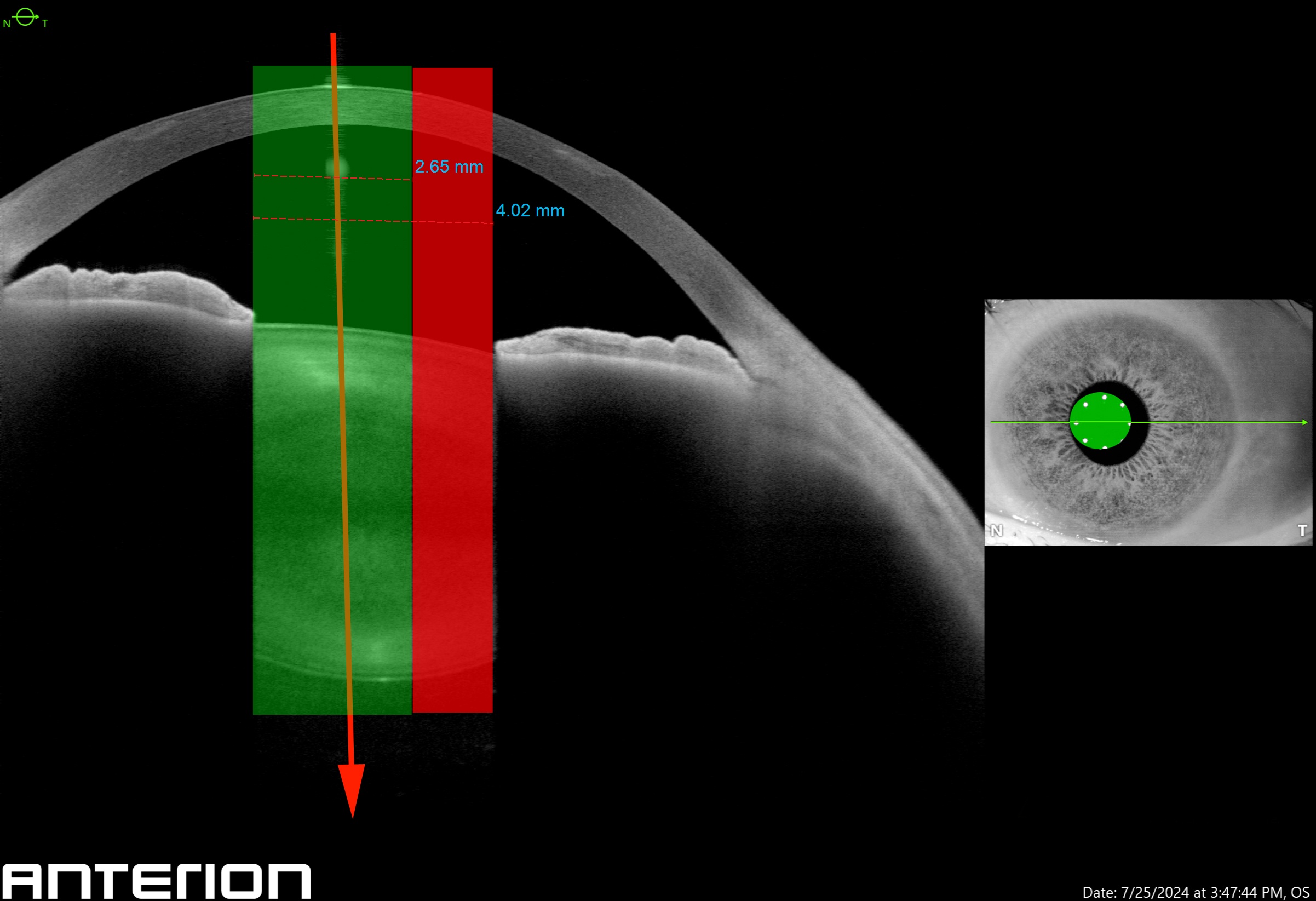
This beautiful image is from our newest piece of equipment, the Heidelberg Anterion. It is the most advanced OCT available for the anterior segment, and just approved by the FDA in 2024. This patient’s eye has a large angle Kappa. The eye is slightly rotated so that the light, represented by the red arrow, enters the cornea and passes through the nasal part of the pupil. Since every aberrometer takes a radial or circular scan, we can simulate that scan with the green cylinder. The red indicates the portion of the pupil outside of the measurement. The area of the scan is measured at 5.5 square millimeters. The entire pupillary area is 12.5 square millimeters, which leaves a lot of area not covered by the wavefront scan. The area of the scan is also shown on the infrared inset image. Get the picture now? But there is another reason: when a scleral contact is placed on the eye, the lens itself may cause a displacement of the corneal reflex. It therefore may not really represent the line-of-sight.
Which Is Best?
Although long debated, the line-of-sight method option is being made available by many instrument manufacturers, LASIK surgeons and even contact lens manufacturers. We still prefer the pupil center as the location of the center of the scan and believe it is more reliable. Make sure you know which method is being used by your contact lens provider. You may be surprised that you did not achieve the night vision you were dreaming about.